Celogics

Unlock the Potential of Cardiac Research with High Quality iPSC-Derived Cardiomyocytes
Celo.Cardiomyocytes are Suitable for 2D and 3D Formats and Have Been Used in Numerous Discovery and Safety Applications
Designed for both discovery and safety/toxicity assessment applications, Celogics’ Celo.Cardiomyocytes harness the power of induced Pluripotent Stem Cells (iPSCs) – under serum-free conditions using proprietary protocols – to revolutionize the way we study and understand the intricacies of the human heart. Validated on multiple commercially available platforms, these iPSC-derived cardiomyocytes offer an unparalleled advantage and provide a reliable and physiologically relevant model for studying cardiac function and disease mechanisms.
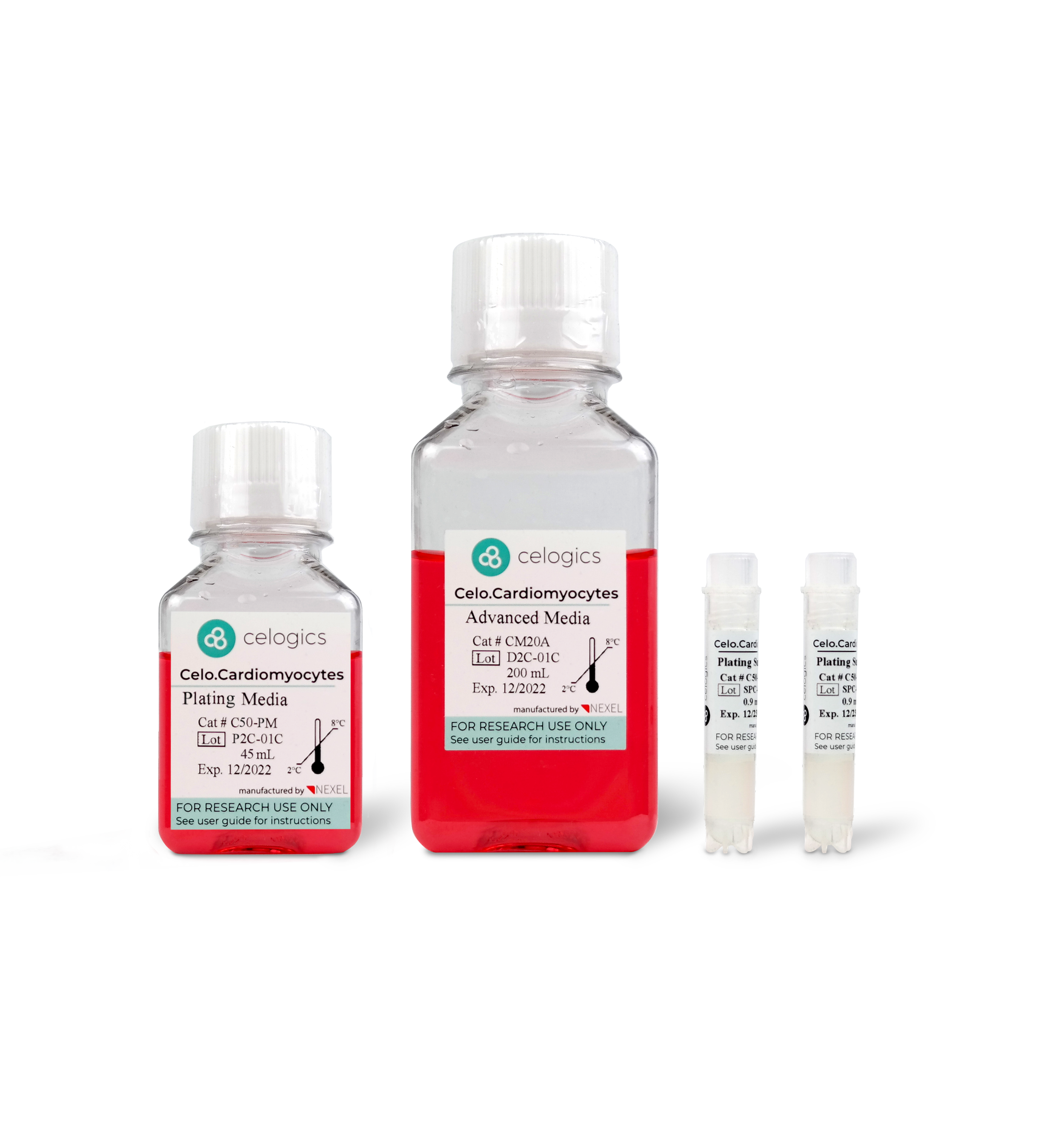
Celo.Cardiomyocytes Products
Celogics recommends using Celo.Cardiomyocytes with their specifically validated media to achieve the best possible results.
Comprehensive Set
$1,300.00
Celo.Cardiomyocytes (5M Cells)
Advanced Media (200mL)
Maintenance Supplement
Plating Media (45mL)
Plating Supplement
Quantity:


Full Media Kit
$200.00
Advanced Media (200mL)
Maintenance Supplement
Plating Media (45mL)
Plating Supplement
Quantity:
Quality Cells
ISO 9001-certified Quality Control
Validated extensively in peer-reviewed journals, application notes, and commercially available platforms.
Electrophysiology-focused product development.
Quality Research
Reproducible Results (low variability)
High Sensitivity to electrophysiological changes from drug treatment.
Serum-free media prevents unwanted interactions & increases biological relevance.
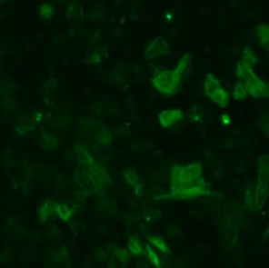
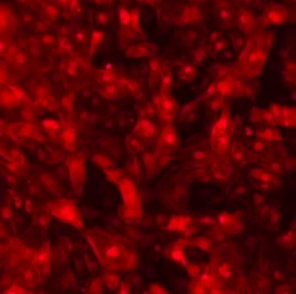
Celo.Cardiomyocytes Morphology
Celo.Cardiomyocytes recover rapidly upon thawing and beat spontaneously from day 2. The beating stabilizes over time (45~60 bpm on DIV7). Users can benefit from a single synchronous monolater formed by these cardiomyocytes which enhances both the reproducibility and accuracy of assays.
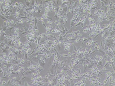
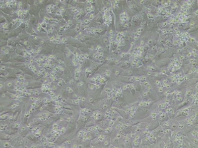
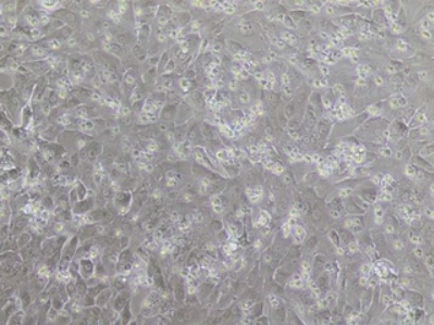
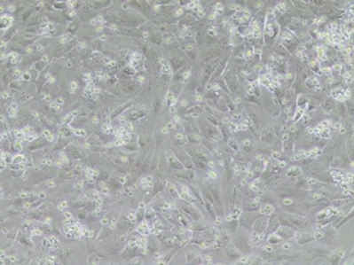
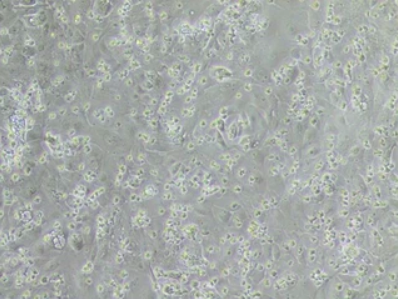
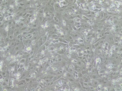
Above, Seeded Celo.Cardiomyocytes on a 12-well culture plate throughout day 1 to 14 post-thawing (magnification 100x) at a plating density of 150,000 cells/cm².
Celo.Cardiomyocytes Express Ventricular Cardiomyocyte-specific Markers
Fluorescence Staining
Celo.Cardiomyocytes express typical ventricular cardiomyocyte markers and structural characteristics. In particular, the Celo.Cardiomyocytes demonstrate high expression of Connexin 43, a gap junction protein, which suggests high interconnectivity and synchronicity.
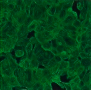
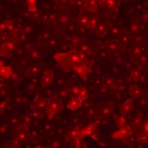
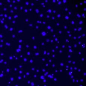
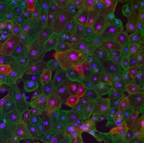

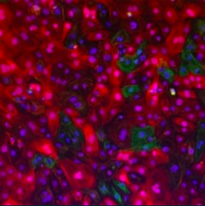
Above, Fluorescence microscopy images of Celo.Cardiomyocytes seeded on Matrigel-coated coverslip at a density of 30,000 cells. Cells were cultured and fixed on day 7.
Celo.Cardiomyocytes Form Robust 3D Engineered Heart Tissues
Combined with Curi Bio’s proprietary serum-free medium kit, Celo.Cardiomyocytes demonstrate robust performance in 3D tissue applications and are ideal for organoid formation and function.


Above, three different Celo.Cardiomyocytes lots were compared in EHTs across one month of culture. As contractile forces continue to increase > 1 month in culture, beating rates continue to slow, suggesting a continuously maturing model.

Celo.Cardiomyocytes Brochure
Download the Celo.Cardiomyocytes product brochure PDF for general information. For additional information, please contact contact@curibio.com.